

17 Aug 2023
The anatomy of bee stingers could help lead to advancements in the emerging field of micro medical devices.
New research deconstructing the anatomy of a honeybee stinger could help pave the way for a future generation of miniaturised medical devices used for drug delivery in humans.
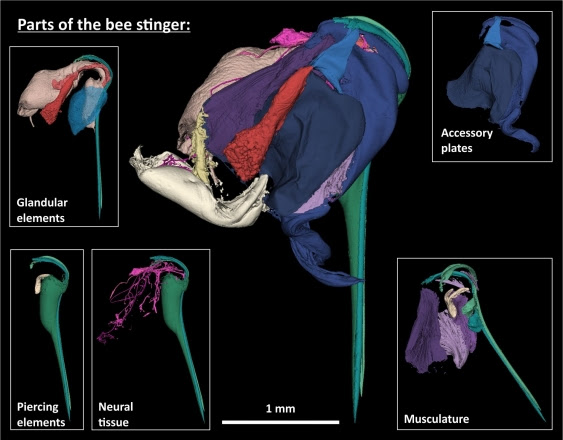
Recently published in the journal iScience, the high-resolution 3D deconstructions produced by UNSW Canberra researchers reveal the unique properties of the bee’s powerful defence mechanism, including the numerous barbs responsible for why the stinger is able to work its way deeper into the skin while pumping venom after stinging.
According to lead researcher, Associate Professor Sridhar Ravi, the autonomous delivery mechanism of the bee stinger has numerous characteristics that could help researchers develop small-scale and minimally intrusive medical devices in the future.
“We have never before produced images with this level of detail, and they have given us tremendous new insights into the functions of the bee stinger,” A/Prof. Ravi said. “Because of these clearer and more precise images, we have uncovered potential opportunities in medical micro drilling, micro-pumps and much more targeted drug delivery.”
A/Prof. Ravi said there is also the possibility of developing improved ‘anchoring’ methods that will allow medical devices or adhesive patches to hold onto the skin without the need for chemical adhesives which can cause irritation or be unviable on moist surfaces, like the inside of the body.
“Previous studies have shown that a bee stinger is very good at piercing skin with minimal force, but it is very hard to remove once it is embedded,” A/Prof. Ravi said. “This is a really useful property for medical devices that need to be very precisely inserted without damaging surrounding tissues.”
The 3D deconstructions have also led to the UNSW Canberra research team developing prototype devices that simulate a bee stinger’s unique piercing and pumping actions.
“A bee’s stinger must be able to firstly pierce skin without buckling, and it must safely detach and coordinate the muscular contractions that generate stinging,” said Dr Fiorella Ramirez Esquivel, the project’s other primary researcher. “This means both working itself deeper into tissue and pumping venom quickly and efficiently.”
Dr Ramirez Esquivel said because a bee stinger is so small – just approximately 2mm in length – the research team had to use a combination of techniques to observe the stinger and decode how it works.
“[The 3D de-constructions] have been fantastic because they allowed us to 3D print the whole stinger and blow it up to a scale where we can move all the parts around to figure out how they work together,” Dr Ramirez Esquivel said. “High-speed filming the stinger in action was also a significant challenge, but it has been instrumental in understanding how it functions.”
Dr Ramirez Esquivel said that understanding the evolution of the bee’s stinger is a great example of how we can make progress by learning more about other animal and plant species.
“Bee stingers are incredibly complex structures with numerous moving components that also happen to be incredibly effective and efficient at what they do,” Dr Ramirez Esquivel said. “The more we look into it, the more we find amazing intricacies related to how it does its job.”
The researchers say they are excited by the potential of different bio-inspired designs in medicine.
“As advanced manufacturing makes strides in what it is possible for us to make, natural materials like the insect cuticle will become more and more relevant to the design of soft robots and microdevices,” Dr Ramirez Esquivel said.